19 Nisan 2013 Cuma

What Is Heart Disease?


Cardiovascular disease includes a number of conditions affecting the structures or function of the heart. They can include:
  • Coronary artery disease (narrowing of the arteries)
  • Heart attack
  • Abnormal heart rhythms or arrythmias
  • Heart failure
  • Heart valve disease
  • Congenital heart disease
  • Heart muscle disease (cardiomyopathy)
  • Pericardial disease
  • Aorta disease and Marfan syndrome
  • Vascular disease (blood vessel disease)
diseases affecting heart and cardiovascular system
Cardiovascular disease is the leading cause of death for both men and women in the U.S. It is important to learn about your heart to help prevent heart disease. And, if you have cardiovascular disease, you can live a healthier, more active life by learning about your disease and treatments and by becoming an active participant in your care.

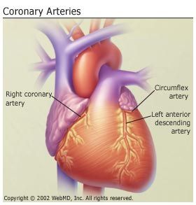
Coronary Artery Disease

Coronary artery disease (CAD) is atherosclerosis, or hardening, of the arteries that provide vital oxygen and nutrients to the heart.

Abnormal Heart Rhythms

The heart is an amazing organ. It beats in a steady, even rhythm, about 60 to 100 times each minute (that's about 100,000 times each day!). But, sometimes your heart gets out of rhythm. An irregular or abnormal heartbeat is called an arrhythmia. An arrhythmia (also called a dysrhythmia) can involve a change in the rhythm, producing an uneven heartbeat, or a change in the rate, causing a very slow or very fast heartbeat.

Heart Failure

The term "heart failure" can be frightening. It does not mean the heart has "failed" or stopped working. It means the heart does not pump as well as it should. This then leads to salt and water retention, causing swelling and shortness of breath.  The swelling and shortness of breath are the primary symptoms of heart failure.
Heart failure is a major health problem in the U.S., affecting nearly 5 million Americans. About 550,000 people are diagnosed with heart failure each year. It is the leading cause of hospitalization in people older than 65.

Heart Valve Disease

diseases affecting heart and cardiovascular system
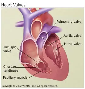
Your heart valves lie at the exit of each of your four heart chambers and maintain one-way blood-flow through your heart.
Examples of heart valve problems include mitral valve prolapse, aortic stenosis, and mitral valve insufficiency.

Congenital Heart Disease

Congenital heart disease is a type of defect in one or more structures of the heart or blood vessels that occurs before birth.
It affects about eight out of every 1,000 children. Congenital heart defects may produce symptoms at birth, during childhood, and sometimes not until adulthood.
In most cases scientists don't know why they occur. Heredity may play a role, as well as exposure to the fetus during pregnancy to certain viral infections, alcohol, or drugs.

Cardiomyopathies

Cardiomyopathies are diseases of the heart muscle itself. People with cardiomyopathies -- sometimes called an enlarged heart -- have hearts that are abnormally enlarged, thickened, and/or stiffened. As a result, the heart's ability to pump blood is weakened. Without treatment, cardiomyopathies worsen over time and often lead to heart failure and abnormal heart rhythms.

Pericarditis

Pericarditis is inflammation of the lining that surrounds the heart. It is a rare condition that is often caused by an infection.

Aorta Disease and Marfan Syndrome

The aorta is the large artery that leaves the heart and provides oxygen-rich blood throughout the body. These diseases and conditions can cause the aorta to dilate (widen) or dissect (tear), increasing the risk for future life-threatening events, such as:
  • Atherosclerosis (hardening of the arteries)
  • High blood pressure
  • Genetic conditions such as Marfan Syndrome
  • Connective tissue disorders (that affect the strength of the blood vessel walls) such as, scleroderma, osteogenesis imperfecta, polycystic kidney disease, and Turner's syndrome
  • Injury
People with aorta disease should be treated by an experienced team of cardiovascular specialists and surgeons.

Other Vascular Diseases

Your circulatory system is the system of blood vessels that carry blood to every part of your body.
Vascular disease includes any condition that affects your circulatory system. These include diseases of the arteries and blood flow to the brain.

What Is Heart Disease?- Heart Palpitations


Heart palpitations are a feeling that your heart is beating too hard or too fast, skipping a beat, or fluttering. You may notice heart palpitations in your chest, throat, or neck.
Heart palpitations can be bothersome or frightening. They usually aren't serious or harmful, though, and often go away on their own. Most of the time, they're related tostress and anxiety or to consumption of stimulants such as caffeine, nicotine, or alcohol. Palpitations also often occur during pregnancy.In about one out of seven cases, the cause can't be identified.
In rare cases, palpitations can be a sign of a more serious heart condition. Therefore, if you have heart palpitations, make arrangements to see your doctor. And seek immediate medical attention if along with palpitations you experience shortness of breath, dizziness,chest pain, or fainting.
After taking your medical history and conducting a physical exam, your doctor may order tests that can either confirm or rule out an underlying cause. If an underlying cause is found, the right treatment can reduce or eliminate palpitations. If your palpitations are not related to an underlying cause, lifestyle changes, including stress management and the avoidance of common triggers, can help prevent them.

Causes of Heart Palpitations

Many things can cause heart palpitations. In the vast majority of cases, the cause is either related to your heart or is unknown. Non-heart-related causes of palpitations include:
Some people experience palpitations after eating heavy meals that are rich in carbohydrates, sugar, or fat. Sometimes eating foods with high levels of monosodium glutamate (MSG), nitrates, or sodium can bring them on.
If you have heart palpitations after eating certain foods, the problem could be food sensitivity. Keeping a food diary can help you identify which foods to avoid.
Palpitations can also be related to underlying heart disease. When they are, palpitations are more likely to represent arrhythmia. Heart conditions associated with palpitations include:

Assessment of Heart Palpitations

Your doctor will conduct a physical examination, take your medical history, and ask about your current medications, diet,and lifestyle. The doctor also will ask when, how often, and under what circumstances palpitations occur.

Assessment of Heart Palpitations continued...

Sometimes, a blood test can reveal the presence of anemia, electrolyte problems, or thyroid abnormalities and help identify the cause of palpitations. Other useful tests include:
  • Electrocardiogram (ECG). An ECG can be done either while you are at rest or while you are exercising. The latter is called a stress ECG. An ECG records your heart's electrical signals and can detect abnormalities in the heart's rhythm.
  • Holter monitoring. A Holter monitor is worn on the chest. It continuously records your heart's electrical signals for 24 to 48 hours. It can detect rhythm abnormalities that weren't identified during a regular ECG test.
  • Event recording.An event recorder is worn on the chest. You use a handheld device to record the heart's electrical signals when symptoms occur.
  • Chest X-ray.
  • Echocardiogram. This is an ultrasound examination of the heart. It provides detailed information about the heart's structure and function.
If necessary, your doctor may refer you to a cardiologist for additional tests or treatment.

Treatment of Heart Palpitations

Treatment of heart palpitations depends on their cause. In most cases, palpitations are found to be harmless and often go away on their own. In those cases, no treatment is needed.
If palpitations are not due to an underlying condition, your doctor may advise you to avoid the things that trigger them. Strategies may include:
  • Reducing anxiety and stress. Common stress-reduction therapies include relaxation exercises, yogatai chibiofeedback, guided imagery, andaromatherapy.
  • Avoiding certain foods, beverages, and substances. This may include alcohol, nicotine, caffeine, and illegal drugs.
  • Avoiding medications that act as stimulants. These include cough and cold medicines, and certain herbal and nutritional supplements.
If lifestyle changes fail to reduce or eliminate  palpitations, your doctor may prescribe certain medications. In some cases, beta-blockers or calcium-channel blockers are used.
If your doctor finds that your palpitations are related to an underlying condition, such as anemia, the focus will be on treating that condition. If the palpitations are caused by a medication your doctor will try to find another medication you can use. If the palpitations represent an arrhythmia, medications or procedures may be required. You may also be referred to a heart rhythm specialist known as an electrophysiologist.

10 Nisan 2013 Çarşamba

What Is Heart Disease?- How the Heart Works


The heart is an amazing organ. It continuously pumps oxygen and nutrient-rich blood throughout the body to sustain life. This fist-sized powerhouse beats (expands and contracts) 100,000 times per day, pumping five or six quarts of blood each minute, or about 2,000 gallons per day.

How Does Blood Travel Through the Heart?

As the heart beats, it pumps blood through a system of blood vessels, called the circulatory system. The vessels are elastic, muscular tubes that carry blood to every part of the body.
Blood is essential. In addition to carrying fresh oxygen from the lungs and nutrients to the body's tissues, it also takes the body's waste products, including carbon ddioxie, away from the tissues. This is necessary to sustain life and promote the health of all parts of the body.
There are three main types of blood vessels:
  • Arteries. They begin with the aorta, the large artery leaving the heart. Arteries carry oxygen-rich blood away from the heart to all of the body's tissues. They branch several times, becoming smaller and smaller as they carry blood further from the heart and into organs.
  • Capillaries. These are small, thin blood vessels that connect the arteries and the veins. Their thin walls allow oxygen, nutrients, carbon dioxide, and other waste products to pass to and from our organ's cells.
  • Veins. These are blood vessels that take blood back to the heart; this blood has lower oxygen content and is rich in waste products that are to be excreted or removed from the body. Veins become larger and larger as they get closer to the heart. The superior vena cava is the large vein that brings blood from the head and arms to the heart, and the inferior vena cava brings blood from the abdomenand legs into the heart.
This vast system of blood vessels -- arteries, veins, and capillaries -- is over 60,000 miles long. That's long enough to go around the world more than twice!
Blood flows continuously through your body's blood vessels. Your heart is the pump that makes it all possible.

Where Is Your Heart and What Does It Look Like?

The heart is located under the rib cage, slightly to the left of your breastbone (sternum) and between your lungs.
Looking at the outside of the heart, you can see that the heart is made of muscle. The strong muscular walls contract (squeeze), pumping blood to the rest of the body. On the surface of the heart, there are coronary arteries, which supply oxygen-rich blood to the heart muscle itself. The major blood vessels that enter the heart are the superior vena cava, the inferior vena cava, and the pulmonary veins.The pulmonary artery and the aorta exit the heart and carry oxygen-rich blood to the rest of the body.

Where Is Your Heart and What Does It Look Like? continued...

On the inside, the heart is a four-chambered, hollow organ. It is divided into the left and right side by a muscular wall called the septum. The right and left sides of the heart are further divided into two top chambers called the atria, which receive blood from the veins, and two bottom chambers called ventricles, which pump blood into the arteries.
The atria and ventricles work together, contracting and relaxing to pump blood out of the heart. As blood leaves each chamber of the heart, it passes through a valve. There are four heart valves within the heart:
  • Mitral valve
  • Tricuspid valve
  • Aortic valve
  • Pulmonic valve 
The tricuspid and mitral valves lie between the atria and ventricles. The aortic and pulmonic valves lie between the ventricles and the major blood vessels leaving the heart.
The heart valves work the same way as one-way valves in the plumbing of your home. They prevent blood from flowing in the wrong direction.
Each valve has a set of flaps, called leaflets or cusps. The mitral valve has two leaflets; the others have three. The leaflets are attached to and supported by a ring of tough, fibrous tissue called the annulus. The annulus helps to maintain the proper shape of the valve.
The leaflets of the mitral and tricuspid valves are also supported by tough, fibrous strings called chordae tendineae. These are similar to the strings supporting a parachute. They extend from the valve leaflets to small muscles, called papillary muscles, which are part of the inside walls of the ventricles.

How Does Blood Flow Through the Heart?

The right and left sides of the heart work together. The pattern described below is repeated over and over, causing blood to flow continuously to the heart, lungs, and body.

Right Side of the Heart

  • Blood enters the heart through two large veins, the inferior and superior vena cava, emptying oxygen-poor blood from the body into the right atrium of the heart.
  • As the atrium contracts, blood flows from your right atrium into your right ventricle through the open tricuspid valve.
  • When the ventricle is full, the tricuspid valve shuts. This prevents blood from flowing backward into the atria while the ventricle contracts.
  • As the ventricle contracts, blood leaves the heart through the pulmonic valve, into the pulmonary artery and to the lungs where it is oxygenated.

Left Side of the Heart

  • The pulmonary vein empties oxygen-rich blood from the lungs into the left atrium of the heart.
  • As the atrium contracts, blood flows from your left atrium into your left ventricle through the open mitral valve.
  • When the ventricle is full, the mitral valve shuts. This prevents blood from flowing backward into the atrium while the ventricle contracts.
  • As the ventricle contracts, blood leaves the heart through the aortic valve, into the aorta and to the body.

    How Does Blood Flow Through Your Lungs?

    Once blood travels through the pulmonic valve, it enters your lungs. This is called the pulmonary circulation. From your pulmonic valve, blood travels to the pulmonary artery to tiny capillary vessels in the lungs.
    Here, oxygen travels from the tiny air sacs in the lungs, through the walls of the capillaries, into the blood. At the same time, carbon dioxide, a waste product ofmetabolism, passes from the blood into the air sacs. Carbon dioxide leaves the body when you exhale. Once the blood is purified and oxygenated, it travels back to the left atrium through the pulmonary veins.

    What Are the Coronary Arteries of the Heart?

    Like all organs, your heart is made of tissue that requires a supply of oxygen and nutrients. Although its chambers are full of blood, the heart receives no nourishment from this blood. The heart receives its own supply of blood from a network of arteries, called the coronary arteries.
    Two major coronary arteries branch off from the aorta near the point where the aorta and the left ventricle meet:
    • Right coronary artery supplies the right atrium and right ventricle with blood. It branches into the posterior descending artery, which supplies the bottom portion of the left ventricle and back of the septum with blood.
    • Left main coronary artery branches into the circumflex artery and the left anterior descending artery. The circumflex artery supplies blood to the left atrium, side and back of the left ventricle, and the left anterior descending artery supplies the front and bottom of the left ventricle and the front of the septum with blood.
    These arteries and their branches supply all parts of the heart muscle with blood.
    Coronary artery disease occurs when plaque builds up in the coronary arteries and prevents the heart from getting the enriched blood it needs. If this happens, a network of tiny blood vessels in the heart that aren't usually open called collateral vessels may enlarge and become active. This allows blood to flow around the blocked artery to the heart muscle, protecting the heart tissue from injury.

    How Does the Heart Beat?

    The atria and ventricles work together, alternately contracting and relaxing to pump blood through your heart. The electrical system of the heart is the power source that makes this possible.
    Your heartbeat is triggered by electrical impulses that travel down a special pathway through the heart.
    • The impulse starts in a small bundle of specialized cells called the SA node (sinoatrial node), located in the right atrium. This node is known as the heart's natural pacemaker. The electrical activity spreads through the walls of the atria and causes them to contract.
    • A cluster of cells in the center of the heart between the atria and ventricles, the AV node (atrioventricular node) is like a gate that slows the electrical signal before it enters the ventricles. This delay gives the atria time to contract before the ventricles do.
    • The His-Purkinje network is a pathway of fibers that sends the impulse to the muscular walls of the ventricles, causing them to contract.
    At rest, a normal heart beats around 50 to 99 times a minute. Exercise, emotions,fever, and some medications can cause your heart to beat faster, sometimes to well over 100 beats per minute.